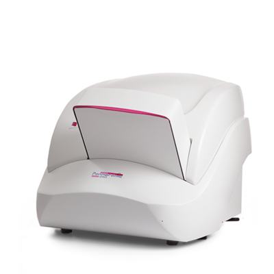
Pannoramic MIDI II
Pannoramic MIDI II – The multi-purpose device for low-level throughput
- Ideal for wet slides due to horizontal feed
- Available with integrated fluorescence unit (optional)
- Automatic tissue detection and scanning
The Pannoramic MIDI II has a capacity of 12 slides and enables you to scan your tissue sections using both bright-field microscopy and a range of fluorescence staining techniques. This makes it a particularly attractive choice for research applications. The tissue sections are loaded horizontally, meaning that wet slides in particular can be scanned without complications.
Speed
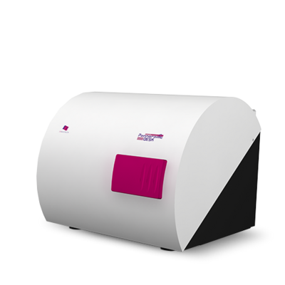
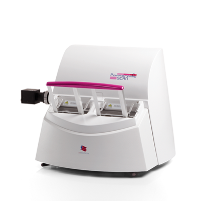
With a scan speed of 90 seconds per slide, the Pannoramic DESK II, MIDI II and SCAN II slide scanners are amongst the fastest on the market.
Quality
All Pannoramic slide scanners provide unique image quality. This is achieved through effective synergy between the camera, the objectives (20x or 40x) and the modern software, which – depending on the camera configuration – provides up to 110x resolution (interpolated) for bright-field and up to 90x resolution (interpolated) for fluorescence microscopy.
Software
Pannoramic control software – included
The Pannoramic control software is the user interface to control your Pannoramic slide scanner. This software supports your daily work processes and the handling of the Pannoramic MIDI II.
To optimise the workflow, all scanners automatically detect the specimen, focus on the tissue and create a preview on the screen. A photograph of the slide label is also created or its barcode is read. This supports archiving, saves time and reduces costs.
- Easy to use
- Choice of manual or automatic scan mode
Automatic tissue detection - Automatic focus
- Photograph of the slide label for documentation
- Optional barcode detection
CaseViewer – included
The CaseViewer is pre-installed on the computer linked to the slide scanner. This software not only allows you to view the digitised tissue specimen, but also offers a wide range of processing options. You can create markings, add comments and view several tissue specimens in parallel in different stainings, to name but a few of the features. To put it simply, the CaseViewer is your gateway to the world of virtual pathology.
| Technology | Bright-field/9-channel fluorescence Objectives: 20x or 40x (optional) Barcode scan: 1D and 2D Bright-field illumination: 3-channel LED Fluorescence illumination: Lumencor light engine Certification: CE, IVD |
| Resolution and magnification | 5-MP basic high-performance CMOS (bright-field and fluorescence): 4.2-MP scientific CMOS (bright-field and fluorescence): |
| Capacity | Slide capacity: 12 Accepted slide formats: 25 x 75 mm, 0.9 to 1.2 mm thickness |
| Digital storage | 3DHISTECH format (MRSX), can be coded for JPG, JPG2000 |
| Scan speed 15 mm x 15 mm (bright-field, 20x objective) | 0.27 µm / pixel (0.63x adapter, 5-MP CMOS): 2 min 30 sec 0.17 µm / pixel (1.0x adapter, 5-MP CMOS): 5 min 30 sec 0.27 µm / pixel (1.0x adapter, 4.2-MP scientific CMOS): 4 min |
| Dimensions (W x D x H) | Pannoramic MIDI II: approx. 52 cm x 57 cm x 42 cm Computer: approx. 20.5 cm x 39 cm x 39.5 cm Mains adapter (24V): approx. 17 cm x 30 cm x 11 cm |
| Weight | Approximately 23 kg |
Sysmex Europe SE
Bornbarch 1
22848 Norderstedt
Germany
+49 (40) 527 26 0
+49 (40) 527 26 100


