Scientific Image Gallery
Welcome to our Scientific Image Gallery. Here you can find real-life examples of cell images, mostly (but not only) from peripheral blood films, that illustrate typical morphologic characteristics pointing to specific conditions or disorders. This constitutes their diagnostic value.
Click on an image to enlarge it and display a short description.

Two lymphocytes of a rabbit. The upper cell has a constricted nucleus which is more often seen with monocytes, but lacks the colour characteristic of monocyte cytoplasm.
<p>Two lymphocytes of a rabbit. The upper cell has a constricted nucleus which is more often seen with monocytes, but lacks the colour characteristic of monocyte cytoplasm.</p>

Monocyte on top, heterophil below. Similar to eosinophils, the granules of leporine neutrophils stain intensely red by May-Giemsa or Wright staining. Therefore, they are termed pseudoeosinophils or heterophils in the rabbit.
<p>Monocyte on top, heterophil below. Similar to eosinophils, the granules of leporine neutrophils stain intensely red by May-Giemsa or Wright staining. Therefore, they are termed pseudoeosinophils or heterophils in the rabbit.</p>

Typical monocyte to the left and tri-segmented heterophil to the right. Similar to eosinophils, the granules of leporine neutrophils stain intensely red by May-Giemsa or Wright staining. Therefore, they are termed pseudoeosinophils or heterophils in the rabbit.
<p>Typical monocyte to the left and tri-segmented heterophil to the right. Similar to eosinophils, the granules of leporine neutrophils stain intensely red by May-Giemsa or Wright staining. Therefore, they are termed pseudoeosinophils or heterophils in the rabbit.</p>

Small lymphocyte with little cytoplasm to the left and a large atypical lymphocyte with abundant, intensely blue cytoplasm to the right.
<p>Small lymphocyte with little cytoplasm to the left and a large atypical lymphocyte with abundant, intensely blue cytoplasm to the right.</p>

Small lymphocyte and a basophil of a rabbit. The basophil granules are well defined and numerous, almost concealing the outline of the nucleus similar to a mast cell.
<p>Small lymphocyte and a basophil of a rabbit. The basophil granules are well defined and numerous, almost concealing the outline of the nucleus similar to a mast cell. </p>

From left to right a band neutrophil, basophil and lymphocyte. In this basophil the basophilic granules are dense, but each granule is blurred, a common finding in rabbit basophils.
<p>From left to right a band neutrophil, basophil and lymphocyte. In this basophil the basophilic granules are dense, but each granule is blurred, a common finding in rabbit basophils.</p>

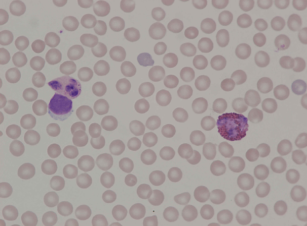
Giemsa simple stained blood smear of a rabbit showing a heterophil (neutrophil) to the upper left, a lymphocyte below and an eosinophil to the right. Note the lighter staining nucleus of the eosinophil as well as a lighter colour of the neutrophilic granules resulting from this stain.
<p>Giemsa simple stained blood smear of a rabbit showing a heterophil (neutrophil) to the upper left, a lymphocyte below and an eosinophil to the right. Note the lighter staining nucleus of the eosinophil as well as a lighter colour of the neutrophilic granules resulting from this stain.</p>

