Scientific Image Gallery
Welcome to our Scientific Image Gallery. Here you can find real-life examples of cell images, mostly (but not only) from peripheral blood films, that illustrate typical morphologic characteristics pointing to specific conditions or disorders. This constitutes their diagnostic value.
Click on an image to enlarge it and display a short description.

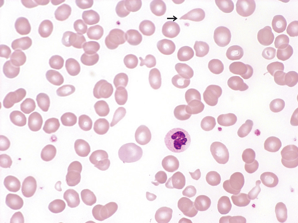
Peripheral blood (May-Grünwald-Giemsa stain) of an 82-year old patient with RCMD showing a macrocytic, hyporegenerative anaemia with clear changes of the red blood cells (micro- and macrocytosis, poikilocytosis and some teardrop cells (->)).
<p>Peripheral blood (May-Grünwald-Giemsa stain) of an 82-year old patient with RCMD showing a macrocytic, hyporegenerative anaemia with clear changes of the red blood cells (micro- and macrocytosis, poikilocytosis and some teardrop cells (->)).</p>

Bone marrow histology (Gomori stain) of a patient showing an infiltration of mantle cell lymphoma cells and an increase in fibres (black lines).
<p>Bone marrow histology (Gomori stain) of a patient showing an infiltration of mantle cell lymphoma cells and an increase in fibres (black lines).</p>

May-Hegglin anomaly belongs to a family of macrothrombocytopenias characterised by mutations in the MYH9 gene. It is a rare autosomal dominant disorder characterised by various degrees of thrombocytopenia that may be associated with purpura and bleeding.
<p>May-Hegglin anomaly belongs to a family of macrothrombocytopenias characterised by mutations in the MYH9 gene. It is a rare autosomal dominant disorder characterised by various degrees of thrombocytopenia that may be associated with purpura and bleeding. </p>

Cell description:
Size: 10-12 µm
Nucleus: kidney or U-shaped with clumped chromatin
Cytoplasm: acidophilic
neutrophil: fine reddish granulation
Cell division is not possible anymore and protein synthesis has stopped.
<p>Cell description: </p> <p>Size: 10-12 µm </p> <p>Nucleus: kidney or U-shaped with clumped chromatin </p> <p>Cytoplasm: acidophilic </p> <p>neutrophil: fine reddish granulation </p> <p>Cell division is not possible anymore and protein synthesis has stopped.</p>

Blood film prepared after storage of the EDTA blood for more than one day. A safe morphological differentiation is no longer possible.
<p>Blood film prepared after storage of the EDTA blood for more than one day. A safe morphological differentiation is no longer possible.</p>

Cell description:
Size: 20 µm
Nucleus: kidney- to band-shaped
Cytoplasm: grey and clear with fine azurophilic granules
They are shortly located in the peripheral blood and then move into the tissue where they differentiate into macrophages. Function: Phagocytosis either of harmful pathogens or dead, dying or damaged cells from the blood.
<p>Cell description: </p> <p>Size: 20 µm </p> <p>Nucleus: kidney- to band-shaped </p> <p>Cytoplasm: grey and clear with fine azurophilic granules </p> <p>They are shortly located in the peripheral blood and then move into the tissue where they differentiate into macrophages. Function: Phagocytosis either of harmful pathogens or dead, dying or damaged cells from the blood. </p>


