Scientific Image Gallery
Welcome to our Scientific Image Gallery. Here you can find real-life examples of cell images, mostly (but not only) from peripheral blood films, that illustrate typical morphologic characteristics pointing to specific conditions or disorders. This constitutes their diagnostic value.
Click on an image to enlarge it and display a short description.

Giant platelets have their name derived from their size exceeding the size of a normal red blood cell.
<p>Giant platelets have their name derived from their size exceeding the size of a normal red blood cell. </p>

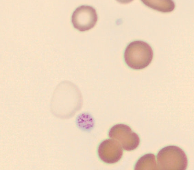
Giant platelet with prominent granulation from a patient with essential thrombocythaemia (ET). (Defective haematopoiesis causes detectable poikilocytosis.)
<p>Giant platelet with prominent granulation from a patient with essential thrombocythaemia (ET). (Defective haematopoiesis causes detectable poikilocytosis.)</p>

Peripheral blood (May-Grünwald-Giemsa stain) of a patient with essential thrombocythaemia, ET. Giant platelets (->) are frequently observed in this disease.
<p>Peripheral blood (May-Grünwald-Giemsa stain) of a patient with essential thrombocythaemia, ET. Giant platelets (->) are frequently observed in this disease.</p>

Granulated blasts have a size of 12 - 16 µm and show significant reddish granules in their cytoplasm. The shape of the nucleus is round or oval with a very high nucleocytoplasmic ratio of 70 – 95%.
The chromatin is predominantly regularly distributed and neither clumped nor condensed. The nucleus has a varying number of nucleoli which may be hidden by the chromatin.
<p>Granulated blasts have a size of 12 - 16 µm and show significant reddish granules in their cytoplasm. The shape of the nucleus is round or oval with a very high nucleocytoplasmic ratio of 70 – 95%. </p> <p>The chromatin is predominantly regularly distributed and neither clumped nor condensed. The nucleus has a varying number of nucleoli which may be hidden by the chromatin. </p>

Granulated blasts have a size of 12 – 16 µm and show significant reddish granules in their cytoplasm. The shape of the nucleus is round or oval with a very high nucleocytoplasmic ratio of 70 – 95%.
The chromatin is predominantly regularly distributed and neither clumped nor condensed. The nucleus has a varying number of nucleoli which may be hidden by the chromatin.
<p>Granulated blasts have a size of 12 – 16 µm and show significant reddish granules in their cytoplasm. The shape of the nucleus is round or oval with a very high nucleocytoplasmic ratio of 70 – 95%. </p> <p>The chromatin is predominantly regularly distributed and neither clumped nor condensed. The nucleus has a varying number of nucleoli which may be hidden by the chromatin. </p>

Normal granulocyte of a healthy individual.
<p>Normal granulocyte of a healthy individual.</p>

Granulocytes with typical reddish granulation of a patient treated with granulocyte growth factor (G-CSF).
<p>Granulocytes with typical reddish granulation of a patient treated with granulocyte growth factor (G-CSF).</p>

Basophils are rarely seen in peripheral blood of guinea pigs. They are only slightly larger than heterophils, have a lobulated homogeneously purple-staining nucleus and numerous round violet granules of varying size.
<p>Basophils are rarely seen in peripheral blood of guinea pigs. They are only slightly larger than heterophils, have a lobulated homogeneously purple-staining nucleus and numerous round violet granules of varying size.</p>