Scientific Calendar June 2024
Presence of unstable haemoglobin
Which scattergram delivers spurious results in the case of a haemoglobin variant?
WNR scattergram
WDF scattergram
RET scattergram
PLT-F scattergram
Congratulations!
That's the correct answer!
Sorry! That´s not completely correct!
Please try again
Sorry! That's not the correct answer!
Please try again
Notice
Please select at least one answer
Scientific background
Variants of the haemoglobin molecule belong to the most common genetic disorders [1]. While the largest portion of the more than 1,200 known haemoglobin variants (Hb variants) have no clinical effect, approximately 150 unstable Hb variants are known to cause haemolytic anaemia of varying severities [2].
On a molecular level, Hb variants are usually caused by single amino acid exchanges in the globin protein leading to different structures and biochemical properties (e.g. oxygen affinity) of the haemoglobin molecule. Mutations resulting in unstable haemoglobin molecules are considered rare events individually. By convention, these variants are named after the geographic origin of the affected individual and most of them are stored in a global gene database. The physiological effects of these Hb variants can range from clinically silent conditions to severe illness [2,3].
Many countries have implemented routine testing of all newborns to identify common haemoglobinopathies. Isoelectric focusing or high-performance liquid chromatography (HPLC) are the most used tests and identify most structurally abnormal haemoglobins. In this way, many benign Hb variants are discovered incidentally [3].
In the current case of the month, we are presenting the measurement results of a patient with the Hb variant ‘Hb G Ferrara’, which was initially described in an Italian family [4]. Since then, it has been described in different case reports as a mildly unstable haemoglobin variant [5 – 6]. In this Hb variant an asparagine residue is replaced by a lysyl residue at position β57 (β57 Asn → Lys) leading to a modification of the structure and properties of the protein and introduction of an extra positive charge that changes the protein’s net charge [4 – 6].
Investigations of the blood smear of patients carrying Hb G Ferrara show altered RBC morphology, such as irregularly shaped RBC and target cells [5].
Case results
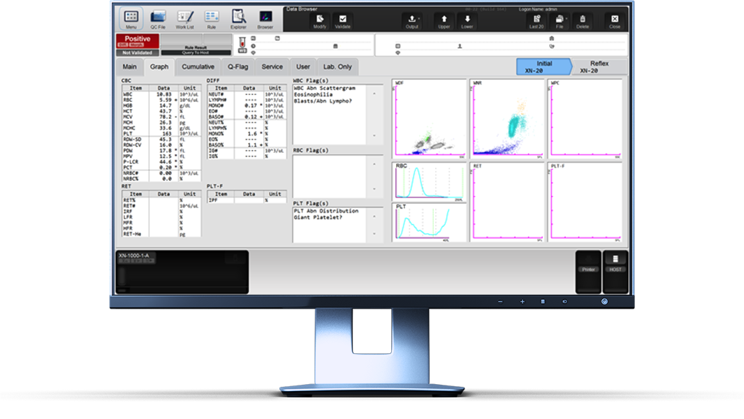
A male patient presented at his general practitioner for a routine check-up. The complete blood count appeared mostly normal, except for a slightly decreased haematocrit and increased RDW-CV triggering the flag message ‘Anisocytosis’. A low MCHC triggered the flag message ‘Hypochromia’.
Interestingly, the WDF scattergram showed all white blood cell (WBC) subpopulations with very low fluorescence signals appearing in the bottom end of the scattergram. Consequently, the XN-Series analyser triggered the ‘WBC Abn Scattergram’ and pointed out the abnormal positioning of the cell clusters.
Scattergram interpretation
It has been observed that the WDF channel analysis of Sysmex XN-Series analysers is affected in the case of haemoglobin abnormalities. It also showed low fluorescence signals that resulted in a low position of the clusters, which led to partially grey cell clusters. However, the WNR channel scattergrams are not affected [7 – 10]. While the cause of this WDF channel alteration is not entirely explained, it is hypothesized that the release of unstable haemoglobin during the RBC lysis might interfere with the WBC staining process by the fluorescent reagent [7]. A possible explanation could be that the polymethine fluorescent marker, used in Sysmex’s haematology analysers for WBC differentiation, might bind to the variant haemoglobin molecule with a greater affinity than to the nucleic acids of WBC [9]. Microscopic observations of affected blood samples have shown no alterations to the WBC populations [8, 9].
Studies have suggested that this pattern distinctly indicates the presence of a haemoglobin variant and should trigger further investigation [7, 8, 10]. Combulazier et al. investigated several patients with different unstable haemoglobin variants and found that the parameters lymphocyte side fluorescence (LY-SFL; equivalent to LY-Y) and neutrophil side fluorescence (NE-SFL) demonstrated excellent sensitivity and specificity. This allowed them to classify 100% of the unstable haemoglobin variant patients in their study cohort [9].
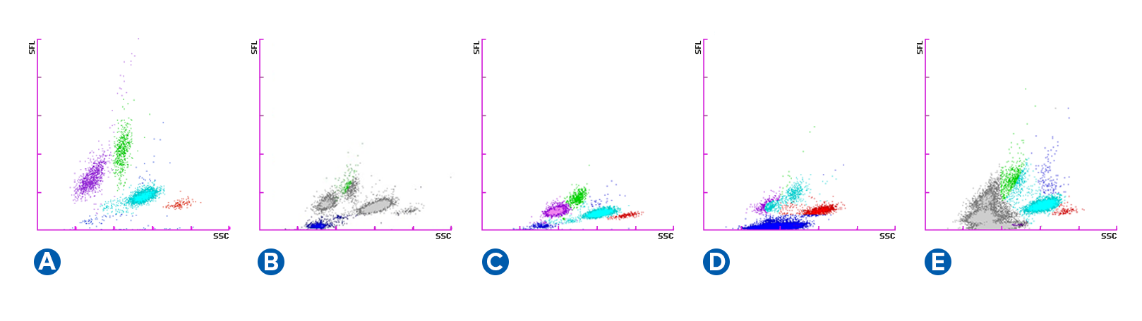
The visible effect in the WDF scattergram can have different intensities as displayed in Fig. 2. A commonality which can be noted is a low fluorescence signal of all cell clusters. Nevertheless, the cluster can still be totally, partially (A and D) or not at all (B and C) greyed out or the cluster analysis can be disrupted (C).
References
[1] Wajcman H et al. (2011): Abnormal haemoglobins: detection & characterization. Indian J Med Res. 134. pp 538 – 546.
[2] Hilbert S et al. (2020): Hemolytic anemia due to the unstable hemoglobin Wien: manifestations and long-term course in the largest pedigree identified to date. Haematologica. 105(5): e253–e255.
[3] Thom CS et al. (2013): Hemoglobin Variants: Biochemical Properties and Clinical Correlates. Cold Spring Harb Perspect Med. 3:a011858.
[4] Giardina B et al. (1978): Properties of hemoglobin G. Ferrara (β57(E1) Asn → Lys). Biochim Biophys Acta. 534(1): 1-6.
[5] Rosetti M et al. (2016): Serendipitous detection of Hemoglobin G-Ferrara variant with Sysmex DIFF channel. Clin Biochem. 49(1-2): 192-3.
[7] Adam AS et al. (2022): Rare unstable and low oxygen affinity haemoglobin variant, Hb Hazebrouck, detected on Sysmex XN-9000. Clin Chem Lab Med; 60(5): e116-e118.
[8] Moioli V et al. (2019): Mozhaisk haemoglobin variant effects on leukocyte differential channel using the Sysmex XN series. Clin Chem Lab Med. 57(12): e324-e327.
[9] Combaluzier S et al. (2023): Detection of unstable haemoglobin variants with Sysmex XN-10. Int J Lab Hematol; 45(2): e21-e23.
[10] Jongbloed W et al. (2018): Unstable haemoglobin variant Hb Leiden is detected on Sysmex XN-Series analysers. Clin Chem Lab Med. 56(9): e249–e250.

It has been extensively described in the literature that samples from patients with unstable haemoglobin variants show a distinct pattern in the WDF scattergram. This SEED article focuses on how users can recognise these special patterns in the scattergrams.

In this case report a blood test was requested for a male patient during a routine check-up, which appeared mostly normal. However, the WDF scattergram presented all WBC subpopulations with very low fluorescence signals appearing in the bottom end of the scattergram and the analyser triggered the flag ‘WBC Abn Scattergram’. A follow-up HPLC analysis revealed the mildly unstable haemoglobin variant ‘Hb G-Ferrara’.